Medical imaging has transformed how doctors diagnose and treat patients. Among the most advanced tools available today is the MRI scanner. This powerful technology creates detailed pictures of the inside of your body without using harmful radiation. Understanding medical imaging helps patients feel more comfortable during their appointments.
How does an MRI work to produce such clear images? The answer lies in the physics of magnets and radio waves. Your body contains millions of water molecules. Each water molecule has hydrogen atoms with protons that spin like tiny magnets. When you enter an MRI machine at facilities like Mayo Clinic or Cleveland Clinic, the strong magnetic field causes these protons to line up in the same direction.
Radio waves then knock these protons out of alignment. When the radio waves stop, the protons snap back into place and release energy signals. The MRI computer captures these signals and turns them into detailed pictures. This process of magnetic resonance imaging explained in simple terms shows why doctors prefer MRI scans for examining the brain, spine, joints, and soft tissues.
MRI technology produces both flat cross-sectional images and 3D pictures of organs and tissues. Doctors use these images to spot problems that other tests might miss. The entire process is painless and safe for most people. Understanding medical imaging and how MRI machines work helps reduce anxiety about this important diagnostic tool.
What is an MRI?
Medical imaging has transformed how doctors diagnose and treat patients. Among the most powerful tools available today, MRI technology stands out for its ability to create incredibly detailed pictures of the human body without using harmful radiation. This non-invasive mri diagnostic procedure has become essential in modern medicine.
Definition of MRI
Magnetic Resonance Imaging (MRI) is a medical imaging technique that uses powerful magnets and radio waves to create detailed pictures of organs and tissues inside the body. Unlike X-rays or CT scans, MRI doesn’t use ionizing radiation, making it safer for repeated use.
The mri machine function relies on the natural magnetic properties of hydrogen atoms in our bodies. Since the human body is mostly water, and water contains hydrogen atoms, MRI scanners can detect these atoms and create images based on their location and concentration.

Overview of MRI Technology
The technology behind MRI is both complex and fascinating. When a patient enters the scanner, the powerful magnetic field aligns hydrogen protons in the body’s tissues. Radio waves are then sent through the body, disrupting this alignment. As the protons return to their original position, they emit signals that the machine captures and converts into detailed images.
This mri diagnostic procedure produces different types of scans, including T1-weighted and T2-weighted images. Each type highlights different tissue characteristics, giving doctors multiple perspectives of the same area. The mri machine function allows physicians to see soft tissues, blood vessels, and organs with exceptional clarity.
The Science Behind MRI
The physics of MRI technology relies on powerful magnetic fields and radio waves working together to create detailed images of the human body. This imaging technique uses the natural properties of hydrogen atoms found in water molecules throughout our tissues. When placed in a strong magnetic field, these atoms behave in predictable ways that allow doctors to see inside the body without surgery or radiation.
How MRI Uses Magnetic Fields
MRI principles start with a powerful magnet that creates a uniform field around the patient. This magnetic field causes hydrogen protons in the body’s water molecules to align in the same direction, like tiny compass needles. The strength of this field typically ranges from 1.5 to 3 Tesla in clinical machines, which is about 30,000 times stronger than Earth’s magnetic field.

Once aligned, these protons spin at a specific frequency called the Larmor frequency. Different tissues contain varying amounts of water, which affects how their protons respond to the magnetic field.
Role of Radio Waves in Imaging
Radio waves pulse through the body at precise frequencies, temporarily knocking the aligned protons out of position. When the radio waves stop, the protons return to their original alignment, releasing energy as radio signals. The physics of MRI technology captures these signals to build images.
Two main timing measurements shape the final image:
- T1 relaxation time measures how quickly protons realign with the magnetic field
- T2 relaxation time tracks how fast protons lose synchronization with each other
Computers use mathematical processes called Fourier transformation to convert these radio signals into the black, white, and gray images doctors examine. Different MRI principles and pulse sequences highlight specific tissues, making various body structures appear brighter or darker for accurate diagnosis.
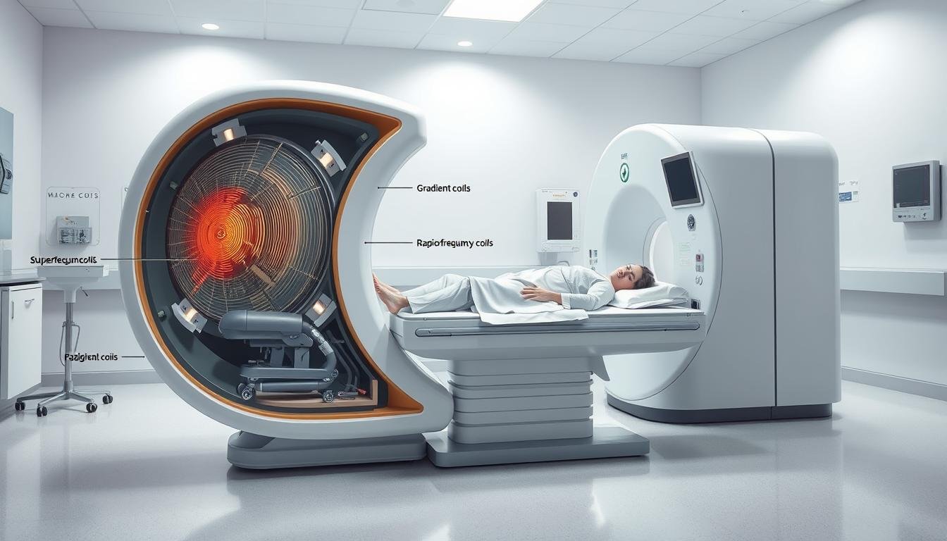
Key Components of an MRI Machine
The MRI scan process relies on three essential components working together to produce detailed images of the body’s internal structures. Each part plays a specific role in how magnetic fields create images that doctors use for diagnosis. Understanding these components helps explain why MRI machines produce such clear pictures of soft tissues, organs, and bones.
Main Magnet
The main magnet forms the heart of every MRI scanner. This powerful component creates a magnetic field up to 60,000 times stronger than Earth’s natural magnetism. The magnet aligns hydrogen protons in your body’s water molecules, preparing them for imaging. Most hospital MRI machines use superconducting magnets cooled to extremely low temperatures—around minus 450 degrees Fahrenheit. This cooling system maintains the magnetic field’s strength throughout the MRI scan process.

Radiofrequency Coils
RF coils work like antennas that both send and receive signals. These specialized devices transmit radio waves into the body and detect the signals that bounce back. Different coils are designed for specific body parts—knee coils, head coils, and spine coils each have unique shapes. The coils sit close to the area being scanned to capture the clearest signals possible.
Gradient Coils
Gradient coils create small variations in the magnetic field that pinpoint exactly where signals come from in your body. These coils produce the loud knocking sounds you hear during a scan. They rapidly switch on and off thousands of times, allowing the machine to build a three-dimensional picture. This precise spatial encoding demonstrates how magnetic fields create images layer by layer.
MRI Procedure Explained
The MRI diagnostic procedure involves careful preparation and specific steps to ensure accurate imaging results. Understanding what happens before and during the scan helps patients feel more comfortable and prepared for their examination.
Preparation for an MRI
Before your MRI scan process begins, you’ll complete safety screening forms about any metal implants or medical devices in your body. The screening is essential because the powerful magnetic field can affect metal objects.
You’ll need to remove all metal items including:
- Jewelry and watches
- Hairpins and eyeglasses
- Hearing aids and dentures
- Underwire bras
- Cosmetics containing metal particles
Staff will provide a hospital gown without metal snaps or zippers to wear during the examination. This preparation ensures your safety and prevents image distortion during the MRI diagnostic procedure.

What to Expect During the Scan
The MRI scan process starts when you lie on a movable table that slides into the tube-shaped scanner. Technologists monitor you from an adjacent room and communicate through a microphone system. You’ll receive a squeeze ball to signal if you need assistance.
Most scans last between 30 to 50 minutes, though some may take 15 minutes to over an hour. The examination consists of multiple scan series with brief pauses between them. Staying completely still during each series prevents image blurring and ensures clear diagnostic results.
Patients who experience claustrophobia can request sedation before the procedure begins. The scanning room remains well-lit and ventilated throughout the entire MRI diagnostic procedure.
Types of MRI Scans
Different MRI scan types serve specific diagnostic purposes in understanding medical imaging. Each technique offers unique advantages for examining various body systems and detecting specific conditions. Medical professionals select the most appropriate scan type based on patient needs and the area being examined.
Functional MRI (fMRI)
Functional MRI measures blood flow changes in the brain during different activities. This advanced form of magnetic resonance imaging explained how brain regions activate when performing tasks like speaking or moving. Doctors use fMRI to map critical brain functions before surgery and detect early signs of neurological conditions like Alzheimer’s disease.

Contrast-Enhanced MRI
Contrast-enhanced scans use gadolinium-based agents injected through an IV catheter. This substance changes the magnetic properties of nearby tissues, making tumors, infections, and blood vessel problems appear clearer on images. The contrast material helps radiologists identify:
- Small tumors or metastases
- Active inflammation areas
- Blood vessel abnormalities
- Infection sites
Open MRI
Open MRI machines feature a two-sided open design instead of the traditional tunnel shape. This configuration benefits patients with claustrophobia or those who cannot fit in standard machines. While open MRI systems may produce slightly lower image quality, they make understanding medical imaging possible for patients who cannot tolerate enclosed spaces. These machines accommodate larger patients and allow parents to stay with children during scans.
Safety and Risks of MRI
Understanding how does an MRI work includes knowing its safety profile. MRI scans are generally very safe medical procedures that don’t use ionizing radiation. The strong magnetic fields used in MRI machine function are not harmful to most people when proper safety guidelines are followed.
Contraindications for MRI
Certain medical implants can interfere with how does an MRI work and pose serious risks. Patients with the following devices typically cannot have MRI scans:
- Pacemakers and older cardiac defibrillators
- Cochlear implants
- Brain aneurysm clips
- Vagal nerve stimulators
- Implanted drug pumps
- Some artificial heart valves

Patients must inform their doctor about any metal in their body. This includes joint replacements, surgical staples, metal pins, screws, plates, stents, bullets, or shrapnel. Pregnancy is typically avoided during MRI scans due to unknown effects on developing babies. People with metal fragments near their eyes need special screening before an MRI.
Potential Risks and Side Effects
The MRI machine function rarely causes problems for most patients. Gadolinium contrast dye, when used, may cause mild allergic reactions in some people. These reactions are usually minor and easily treated with medication. The loud noises during scanning can be uncomfortable but are managed with earplugs or headphones. Some patients experience mild claustrophobia inside the scanner, which technicians can help manage through communication and comfort measures.
Interpreting MRI Results
Reading MRI scans requires specialized training and an understanding of the physics of MRI technology. Radiologists examine different image sequences to identify normal and abnormal tissue patterns. Each scan produces multiple image types that highlight various tissue characteristics based on fundamental MRI principles.
How Radiologists Analyze Images
Radiologists primarily work with T1-weighted and T2-weighted images. These sequences show tissues differently based on their water content and molecular structure. On T1 images, fat appears bright while water appears dark. T2 images reverse this pattern, making fluid-filled areas appear bright.
The physics of MRI technology allows radiologists to spot inflammation, which typically appears dark on T1 images and bright on T2 images. By comparing these different sequences, doctors can determine tissue composition and identify abnormalities with remarkable accuracy.

Commonly Identified Conditions
Different body regions reveal specific conditions through MRI scanning:
- Brain and spine: Aneurysms, tumors, multiple sclerosis, stroke damage, and pinched nerves
- Heart: Chamber abnormalities, valve problems, and congenital defects
- Abdomen and pelvis: Liver cirrhosis, Crohn’s disease, and various tumors
- Bones and joints: Torn ligaments, cartilage damage, and bone infections
- Breast tissue: Cancer detection in dense tissue when combined with mammography
Understanding basic MRI principles helps patients appreciate why certain conditions appear clearly on scans while others require additional imaging sequences or contrast agents for proper diagnosis.
Advances in MRI Technology
Medical imaging continues to evolve at a rapid pace, with MRI technology leading breakthrough discoveries in diagnosis. Recent innovations have transformed the MRI diagnostic procedure into an even more powerful tool for detecting diseases earlier and with greater precision. These advancements build on the fundamental principle of how magnetic fields create images, pushing the boundaries of what doctors can see inside the human body.
Innovations in Imaging Techniques
New imaging methods have revolutionized brain and spinal cord visualization. Diffusion-weighted imaging now detects strokes within minutes by tracking water movement in brain cells. This technique shows restricted water flow in damaged tissue before other signs appear.

FLAIR (Fluid Attenuated Inversion Recovery) sequences separate normal fluid from abnormal tissue with remarkable clarity. Gadolinium contrast agents reveal blood-brain barrier disruptions in conditions like tumors and multiple sclerosis. These tools demonstrate how magnetic fields create images that capture minute anatomical details in three dimensions.
Future of MRI in Medical Diagnosis
Emerging technologies promise faster scans with higher resolution. Artificial intelligence now assists radiologists in spotting subtle abnormalities. Real-time MRI captures moving organs like the beating heart with unprecedented detail.
Scientists are developing portable MRI machines for bedside imaging and emergency situations. Ultra-high field magnets reaching 7 Tesla and beyond reveal brain structures invisible to standard scanners. These innovations ensure the MRI diagnostic procedure will remain central to medical care for decades ahead.
Frequently Asked Questions (FAQs)
Understanding medical imaging procedures can raise many questions for patients preparing for their first scan. The MRI scan process involves specific timeframes and safety considerations that vary from person to person. These common questions help patients know what to expect and whether they qualify for this type of imaging.
How Long Does an MRI Take?
Most MRI scans take between 30 and 50 minutes to complete. Some complex examinations of the brain or spine might take over an hour. The MRI scan process includes multiple imaging sequences with short breaks between each series. Patients need to stay still during each sequence to get clear images. The scanner has open ends that allow medical staff to remove patients quickly if needed during emergencies.
Is MRI Safe for Everyone?
MRI scans are safe for most people since they use magnetic fields instead of radiation. People with certain metal implants like older pacemakers from companies like Medtronic or Boston Scientific cannot have MRI scans unless the devices have MRI certification. Pregnant women can have MRI scans but doctors avoid using gadolinium contrast agents unless the benefits outweigh risks. Patients with kidney disease or liver problems might not receive contrast agents due to processing difficulties.
Understanding medical imaging safety helps patients prepare properly. Mothers who breastfeed should talk with their doctors at facilities like Mayo Clinic or Cleveland Clinic about contrast agent use. Patients with severe claustrophobia might need sedation from an anesthesiologist. Emergency patients on ventilators or other life support equipment cannot use standard MRI machines since the magnetic field interferes with these devices.